A randomly selected set of chest radiographs from cases with WHO-defined clinical pneumonia were collected from the Gambian Pneumococcal Surveillance study and from the multi-country Pneumonia Etiology Research in Child Health (PERCH) study. These were blindly interpreted by each member of the WHO-CRES technical working group using the WHO standardized definitions. Radiographs with at least 66% agreement for the conclusion were collated as a set of reference images for teaching and standardization assessments. Each of these has a descriptive comment explaining the findings present, and a simplified illustration if pathology is present.
Guidelines for the training and assessment of chest radiograph readers and support for studies in the form of a centralised arbitration process for discordant chest radiograph interpretations are currently under development.
These samples images are examples of reference chest X-rays developed as part of the WHO CRES project. These X-rays were read using the WHO standardized methodology and show examples of each WHO category. Annotations and explanations are included for each image. In order to access additional chest X-rays users will need to register on the website and provide some details regarding proposed use of the images. Registration will allow users to access a bank of approximately 450 X-rays with explanations and annotations. In the future additional training material and access to a centralised arbitration process is planned. This will allow registered members to submit images with discordant interpretations as part of research studies to the panel for reading.
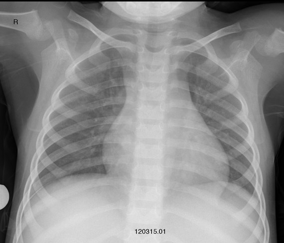
CRES-019
- Quality
- Adequate
- Signif pathology
- No
- Endpoint consolidation (right)
- No
- Endpoint consolidation (left)
- No
- Other infiltrate (right)
- No
- Other infiltrate (left)
- No
- Pleural fluid-R
- No
- Pleural fluid-L
- No
- Silhouette
- No
- WHO Conclusion
- No consolidation/infiltrate/effusion
- Final descriptive comment
- No endpoint consolidation, pleural effusion, or other infiltrate.
This website contains all material to support chest radiograph methods in the design and implementation of an epidemiological study. This includes information and tools for investigators, site staff including radiographers and research staff, and physicians and radiologists who are interpreting radiographs for a study.
Registration is required to access our reference images.